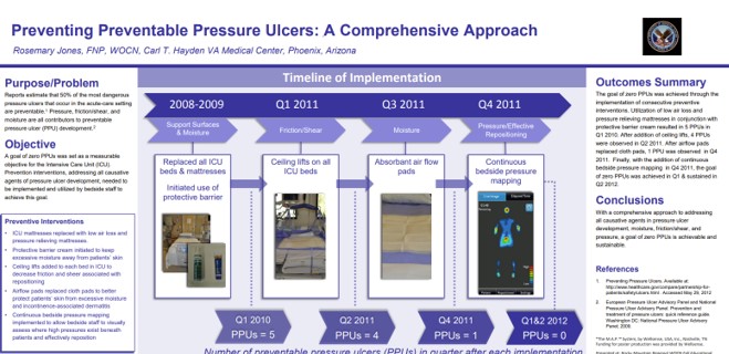
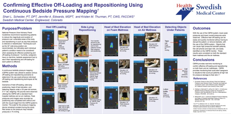
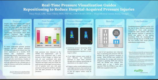
This real-world clinical poster from Floyd Medical Center shows that using a real-time pressure visualization monitor led to a 71% reduction in hospital-acquired pressure injuries (HAPIs) over 5 months in the critical care unit, by guiding staff to reposition patients more effectively and reduce peak pressure exposures.

At the Carl T. Hayden VA Medical Center, a stepwise prevention program that included real-time pressure visualization reduced preventable pressure ulcers (PPUs) from 5 per quarter to 0, maintaining 0 PPUs across 2 consecutive quarters.

Pressure visualization can be used as a structured education tool for appropriate patients, enabling them to learn and reliably perform self-repositioning with clear visual guidance, reducing staff burden while keeping turning technique verifiable.

When appropriate patients were identified as able to self-turn, real-time pressure visualization helped them reposition themselves reliably with clear, immediate feedback. This demonstrates that pressure visualization can actively engage patients in their own pressure management, supporting safer self-directed turning when clinically appropriate.

At Henry Ford Hospital, 101 patients identified as able to self-turn were able to reposition themselves reliably when provided a pressure-visualization system and appropriate instruction, demonstrating that mobile, lower-risk patients can self-manage turning effectively under guided use.

In two UK NHS hospitals, continuous bedside pressure mapping helped identify and reduce pressure ulcer incidence, with staff reporting 100% agreement that the system improved positioning, reduced risk, and was beneficial in care. Patients and families also responded positively, with 78% rating the system helpful.

In a veteran population, pressure visualization was used not only to guide clinical care but to empower patients and caregivers with understandable, visual information about pressure risk. This supports greater patient involvement in day-to-day positioning decisions and reinforces shared accountability for prevention and healing.

SSM DePaul Health Center showed that continuous bedside pressure mapping identified dangerously high sacral pressures at 40–45° head-of-bed elevation, enabled simple micro-shifts to reduce pressure, and guided use of higher support surfaces only when truly needed—improving outcomes and lowering costs

When clinical requirements force elevated head-of-bed, pressure mapping supports safer, repeatable micro-adjustment workflows by showing staff when sacral pressures become excessive and guiding small positioning changes that meaningfully reduce pressure without disrupting the care plan.

Pressure mapping strengthened bedside workflow by allowing staff to visually confirm that a turn actually offloaded pressure, and by helping staff identify and remove hidden causes of high pressure (workflow-relevant problems that are easy to miss during routine care).